- Our clinic
- Magnetic resonance (MRI)
- Services
- Patients
- Physicians
- Contact us
- Make an appointment
- Our clinic
- Magnetic resonance (MRI)
- Services
- Patients
- Physicians
- Contact us
- Make an appointment
![]()
Radiologie générale
- Magnetic resonance (MRI)
- Computed Tomography (CT Scan)
- Positron emission tomography (PET-CT)
- Prostate Imaging
- Breast imaging / Mammography
- Musculoskeletal imaging
- Ultrasound and Doppler
- Virtual colonoscopy
- General radiology
- Infiltrations & Facet joint injections
- Bone densitometry
- Dental imaging
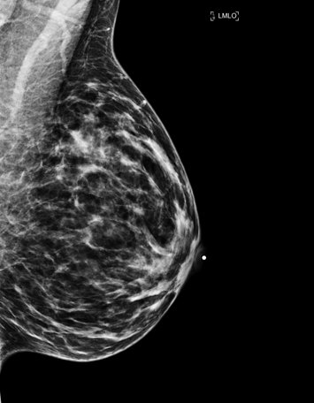
DESCRIPTION (Mammogram)
« RésoScan CLM is a Ministère de la santé et des services sociaux designated screening centre (CDD) for the Québec breast cancer screening program (PQDCS) and is a member of the Canadian Association of Radiologists. »
 By appointment only
Covered by the RAMQ
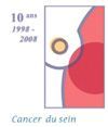
Breast cancer is the type of cancer most often diagnosed in women in Quebec. It is estimated that one in nine women risks developing this cancer during her lifetime. Being a woman and aging are the two main risk factors. Family history of breast cancer (grandmother, mother, sister, etc.) is also a risk factor, but the majority of women with breast cancer do not have a family history of the disease. Even if you do not feel anything, mammography can find very small changes in your breasts.
 A screening mammogram is an X-ray of the breasts to detect small lesions that are often not palpable. Usually, two x-rays are required for each breast: a frontal view and an oblique view.
The breast is compressed to obtain a precise image of the inside of the breast and to reduce the quantity of radiation required by reducing the thickness of the breast. The risk of developing cancer due to repeated mammograms is practically nonexistent.
Digital technology slightly lowers the dose compared to the old analog technology. However, the compression level remains identical and the exam is performed in the same manner. On the other hand, because the images are digital, the total duration of the exam decreases.
Some women experience discomfort or pain when the breast is compressed. This is usually brief and temporary and should not be a cause for concern. If your breasts are sensitive, it is best to avoid consuming coffee, tea, chocolate and soda during the two weeks prior to the exam. For the same reason, it is recommended that the mammogram be done within ten days after the start of your menstrual period or when your breasts are less sensitive.
Mammography is currently recognized as the most effective method to detect breast cancer. In addition, digital technology offers a little more detail than analog technology. However, it also has its limits. Indeed, it is estimated that in about 7% of participants in a breast cancer screening program, the mammography results are interpreted as uncertain.
In the vast majority of these cases, there is no breast cancer. In fact, in the screening context, the majority of exams reveal non-cancerous lesions.
Breast MRI is indicated in two specific situations. First, it can help in the screening of breast cancer, in association with mammography. This is for patients with high risk of breast cancer through their lifetime. It includes patients with BRCA-1 and BRCA-2 gene mutation, as well as their first untested first relative. Also, patients with rare disease like Li-Fraumeni syndrome, Cowden syndrome and Bannayan-Riley-Ruvalcaba syndrome, as well as their first degree relative. Women who underwent chest irradiation between the age of 10 and 30 years can also be screened with breast MR. Finally, any women with a risk of 20% or more as determined with different risk assessment models can also be included. Breast MR also have a place in the evaluation of patient who had a recent diagnosis of breast cancer, especially if it is an invasive lobular carcinoma. Exceptionally, breast MR can be useful in the assessment of the extent of the disease in certain women, helping to plan treatment accordingly.
When a mammogram shows abnormal or uncertain signs, it is necessary to perform further tests to establish an exact diagnosis.
Mammography is an exam used for early detection of breast cancer and diagnosis of other more common breast pathologies. In addition to mammography, we offer breast ultrasound exams when necessary.
Screening mammograms are covered by the RAMQ beginning at age 40:
- with a doctor's prescription for women aged 40 to 49 or age 70 and over;
- with an invitation letter from PQDCS or a doctor's prescription for women aged 50 to 69.
Diagnostic mammograms for women of all ages with a prescription from a doctor are also covered by the RAMQ.
Also offered:
- Breast ultrasound
- Ultrasound-guided cyst puncture
- Ductography
- MRI (magnetic resonance imaging) of the breast
It is possible that after seeing the results of your mammogram, your doctor (or your CDD) will ask you to do further exams. In such a case, this does not necessarily mean you have breast cancer.
There are various types of exams, each corresponding to a specific need. Of course, you will not have to undergo all these tests, only those required in your particular situation.
These additional tests may include :
X-ray images
Additional mammograms to clarify the nature of the lesion.
Exam using ultrasound
Primarily to determine if there are cysts in the breast.
Fine needle biopsy
Cell sampling performed with a fine needle in the location where a lesion is found in the breast.
Core needle biopsy
Tissue sampling with a larger needle in a breast lesion.
Surgical biopsy
Sample of a portion of the lesion through surgery.
Stereotactic localization
Allows placement of a wire in a non-palpable lesion with computer guidance. This wire serves as a guide for surgical removal at the precise area where the lesion is located.